Experimental facilities in the Weeks lab

Located in Emerson Hall 347 & 350 -- see photos of our lab

Lab Home -- People -- Experimental facilities -- Publications -- Experimental pictures -- Links

Capabilities we have

We can do lots of cool things. Our current capabilities (August 2025) include:
  • Regular, brightfield microscopy:
    Magnifications from 1.6x to 160x. (The best resolution we can get is 0.2 microns.) See table below for more information on our lenses. We have both inverted and upright microscopes. We also have a rotation stage: we can rotate the sample in a horizontal plane while we're looking at it.

  • Differential interference contrast (DIC) microscopy:
    This technique is useful for looking at thin samples, or samples with low contrast.

  • Fluorescence microscopy:
    Biologists love this, as they have all sorts of clever ways to make odd things fluoresce. Physicists (like us) like using boring old fluorescent particles.
    • We have a variety of sizes of fluorescent polystyrene particles on hand, typically with sizes around 1 micron diameter.
    • See our links page for sources of these particles.

  • Temperature control:
    We have a temperature control system for the microscope, which works from room temperature up to about 40 degrees C. We also have a water bath which can be used to control temperatures.

  • Microfluidics capabilities:
    We can do soft lithography to design and fabricate microfluidic devices. To an extent, we are using resources available through campus facilities. In our lab we have a spin-coater which is useful for this work.

  • High speed camera:
    We have a Phantom V9.1 camera with 6 GB of memory. This is a 12-bit camera capable of 1632x1200 pixels at 1016 frames per second, and 96x8 pixels at 153,846 frames per second, as well as other ranges in between.

  • Pipette puller:
    We have a pipette puller and a microforge, suitable for making glass pipettes with tip sizes down to a few microns.

  • Analysis:
    We use IDL to analyze our images. We have several Linux computers for this purpose. While Eric prefers IDL, at any given moment in the lab group, there are usually some folks who are using Python and some using Matlab.

  • Particle tracking:
    Our favorite analysis technique is tracking the motion of individual colloidal particles. Best of all, we can do this for several thousand particles simultaneously, in three dimensions.

  • Rheometer:
    We have an old TA instruments rheometer.

  • Particle synthesis:
    In the distant past, we made fluorescent colloidal PMMA; the last batch we made was back in 2011. We still have the capability, in theory.

  • Langmuir trough:
    We have a Kibron Langmuir trough. This has not been used for more than a decade, though.

  • Miscellaneous Gadgets:
    These include a refractometer, several viscometers, and several simple cameras.

  • Obsolete equipment:
    This webpage used to brag about how we could digitize videotapes, and how we have a video enhancement box to improve video signals traveling through coax cables. I'm not sure we could digitize videotapes at this point, and I don't think anybody uses coax cables for video cameras these days.


Microscope objectives that we have

Magnification/NA theoretical
resolution
working
distance
comments
1.6x / 0.05 air6 um3.4 mm-
5x / 0.15 air3.3 um12 mm-
10x / 0.22 air1.3 um5.8 mm-
10x / 0.40 air0.7 um2.2 mm-
10x / 0.40 oil0.7 um0.36 mm-
20x / 0.40 air0.7 um1.9 - 3.2 mm long working distance lens, with a correction collar for glass thicknesses from 0.0 to 2.0 mm
20x / 0.70 multi (oil)0.4 um250 umThis lens can work with a variety of immersion fluids, including water, glycerol, and oil. In general, consider this an oil immersion lens.
40x / 0.55 air0.5 um1.9 - 3.3 mmlong working distance lens, with a correction collar for glass thicknesses from 0.0 to 2.0 mm
40x / 1.25 oil0.23 um100 um-
63x / 0.70 air0.4 um1.8 mm long working distance lens, with a correction collar for glass thicknesses from 0.1 mm thick up to 1.3 mm (in other words, a microscope slide or petri dish).
63x / 1.20 water0.24 um220 umBest lens for aqueous samples, such as most biological samples. This is a really nice lens.
100x / 1.35 oil0.21 um90 um-
100x / 1.40 oil0.20 um90 umThe "highest power" lens. It helps if your samples have an index of refraction similar to oil, in other words, close to n=1.5.

NOTES:


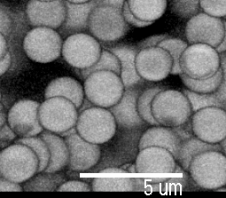
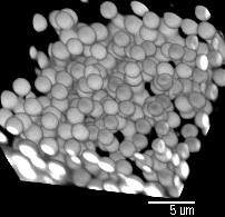
We used to have a confocal microscope that took these 3D pictures:


Pictures by Piotr Habdas, showing colloidal particles which have formed a gel. Another image of the same sample is the background for this web page.