Research
Contents
• Brief overview
• Solvent-protein-reaction dynamical coupling in enzyme catalysis
• Protein nucleation and assembly; creation of novel microenvironments and reactivities
• B12 trafficking and delivery enzymes in humans (in collaboration)
• EPR spectroscopy: Innovations & Applications
Brief Overview
A principal goal of the biophysics research in the Warncke Lab is to elucidate fundamental physical-chemical mechanisms of coupled solvent and protein dynamics that actuate and orchestrate catalysis in enzymes, with a focus on radical catalysis in coenzyme B12 (adenosylcobalamin, AdoCbl) –dependent enzymes.
Aligned with this goal, we also address fundamental features of the processes involved in the non-covalent assembly of proteins to form structures, that lead to emergent reactivities, and that create solution microenvironments with unique structures and dynamics.
The systems we study involve native metal ion and organic radical paramagnetic states, or introduced paramagnetic spin probes and spin labels. Our principal physical techniques of pulsed- and continuous-wave- electron paramagnetic resonance (EPR) spectroscopy detect spatial (tenths to 10’s of Angstroms) and temporal (picoseconds to megaseconds) information from the unpaired electron spins, that reveal fundamental mechanisms of biological function.
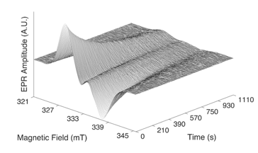
of EAL radical pair EPR spectrum at 210 K.
Following are compact descriptions of principal on-going projects:
Solvent-protein-reaction dynamical coupling in enzyme catalysis
Reactions
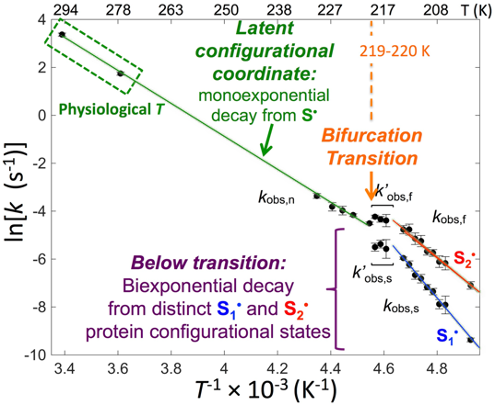
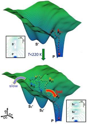
A combination of low temperatures (173-250 K) and aqueous-cryosolvent systems has been used to parse individual steps from the catalytic cycle of the EAL enzyme for first-order kinetics analysis. Time-resolved, full-spectrum EPR reveals the protein configurational states and configurational fluctuations that actuate and guide the core steps in catalysis. The methods provide unprecedented access to the inner workings of an enzyme.Figures 1, 2 and 3 illustrate low-temperature, time-resolved EPR results, Arrhenius plot of fitted reaction rate constants showing reactivity regions, and free energy landscape representation. For details, see: Publications 54, 56, 59.
Solvent Dynamics and Protein-Solvent Coupling
decay reaction pathways and rate-limiting steps.
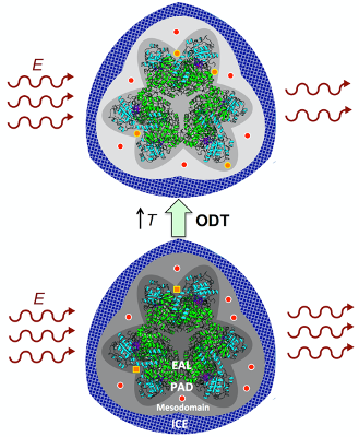
Figure 4 depicts the solvent structure around the EAL enzyme, and the temperature-dependent dynamics, including the order-disorder transition in the PAD, that are detected by the spin probe. Also depicted is a new experimental approach, which directly detects solvent fluctuations through the influence of dielectric permittivity on EPR amplitude.
For details, see: Publications 62, 69.
Putting it Together: Solvent-Protein-Reaction Dynamical Coupling
We are combining the reaction-kinetic and solvent-dynamic studies in the tunable low-temperature, mesodomain system to reveal universal features of the solvent-protein-reaction dynamical coupling to adiabatic, bond-making/bond-breaking steps in enzyme catalysis.Details in new publications, coming in 2021!
Protein-solvent microenvironments and reactivities
experiment, with dynamics reported by spin probe
(circles, squares) and dielectric permittivity.
Protein-solvent interactions and creation of unique phases and reactivities
The interactions of protein with solvent generate local structural and dynamical states that impact enzyme reactions. Interactions among protein and solvent can also create global regions of distinct dynamics, such as in liquid-liquid phase separation (LLPS). The spin probe studies in the low-temperature system are being extended from EAL to other proteins, that represent different classes: globular, hybrid globular+disordered domains and fully, intrinsically-disordered proteins (IDPs). Our aim is to understand global phase dynamics, toward control of protein assembly processes and enzyme reactivity.Bacterial microcompartment
The Eut (ethanolamine utilization) bacterial microcompartment (BMC) is delimited by an ~100 nm diameter, polyhedral protein shell, that encapsulates the enzymes involved in ethanolamine metabolism, including the “signature enzyme,” EAL. Widespread use of the BMC architecture for encapsulating metabolic pathways is evidenced across bacterial phyla. To advance our in vitro EPR experimental and analysis methods toward in vivo studies, we have initiated investigation of the assembly and molecular structure of the Eut BMC from Salmonella typhimurium, by using techniques of molecular biology, in combination with EPR spin-labeling.
amyloid fiber with bound Cu2+.
Amyloid-β protein –derived structures (collaboration with Lynn Laboratory, Dept. of Chemistry, Emory)
The Lynn Laboratory creates novel protein frameworks (fibrils, fibers, ribbons, sheets, tubes) from small peptides, derived from the N-terminus of the amyloid-β protein. We study the binding of paramagnetic copper ions, Cu2+, to these frameworks. A representation of this type of assembly is shown in Figure 5. Current work is focused on characterization of the local and global metal site structure of a nonapeptide composed of residues 13-21 of the amyloid-β protein, which includes two Cu2+–binding histidine imidazole side chains. The redox properties of Cu1+/Cu2+ make these assemblies valuable for energe storage and utilization applications.EPR studies of B12 trafficking and delivery enzymes (collaboration with Banerjee Laboratory, University of Michigan)
EPR spectroscopy: Innovations & Applications
Pulsed-EPR spectrometer
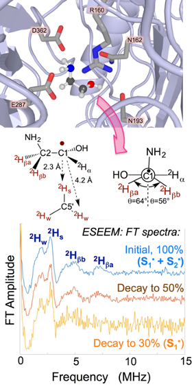
substrate radical. Middle: Depiction of
structure from 3-pulse 2H-ESEEM spectroscopy.
Bottom: 2H-ESEEM of substrate radical
during decay reaction.
We designed, constructed and implemented a pulse programmer, based on the field-programmable gate array (FPGA) technology, for pulsed-EPR experiments. The FPGA pulse programmer offers advantages in design flexibility, cost and speed over digital delay and logic pattern generators, and application-specific integrated circuit (ASIC) devices.
For details, see: Publication 51.
For lab instrumentation, see: Home\Lab\Instrumentation
ESEEM simulation
We developed “OPTESIM” simulation software for the pulsed-EPR technique of ESEEM spectroscopy, within the MATLAB (Mathworks, Natick, MA) environment (Figure 5). Package and tutorials are available for download.For details, see: Publication 44.
For downloads, see: Home\Software
Time-resolved, full-spectrum EPR
We developed two protocols for sample preparation and methods for measurement of kinetics of single enzyme reaction steps, parsed from the catalytic cycle, by using cryosolvents and low temperatures. We described these in the detailed format of a Methods in Enzymology chapter. These experiments utilize the commercial Bruker E500 continuous-wave EPR spectrometer.Time-resolved, full-spectrum EPR spectroscopy data is shown in Figure 1.
For details, see: Publication 54.